Research
1. Microparticle dynamics in acoustofluidics
We develop analytical and numerical models to explain the observed dynamics of microparticles driven by acoustic forces in microfluidic devices.
1.1 Acoustic radiation force and torque on microparticles
Jose P. Leao-Neto, & Glauber T. Silva
Collaboration with ESPCI-Paris
Publications:
- JP Leao-Neto, M Hoyos, JL Aider, GT Silva, Acoustic radiation force and torque on spheroidal particles in an ideal cylindrical chamber The Journal of the Acoustical Society of America 149, 285-295, 2021.
- EB Lima et al., Nonlinear Interaction of Acoustic Waves with a Spheroidal Particle: Radiation Force and Torque Effects, Physical Review Applied 13, 064048, 2020.
-
JH Lopes, EB Lima, JP Leão-Neto, GT Silva, Acoustic spin transfer to a subwavelength spheroidal particle, Physical Review E 101, 043102, 2020.
- JP Leão-Neto, JH Lopes, GT Silva, Acoustic radiation torque exerted on a subwavelength spheroidal particle by a traveling and standing plane wave The Journal of the Acoustical Society of America 147, 2177-2183, 2020.
-
GT Silva et al., Particle patterning by ultrasonic standing waves in a rectangular cavity, Physical Review Applied 11, 054044, 2019.
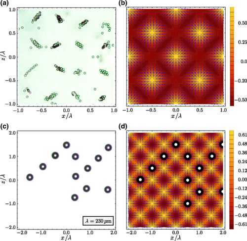
Polystyrene particles with a diameter of (a) 10μm and (c) 75μm in a rectangular acoustofluidic device. (b) and (d) The corresponding theoretical predictions for the radiation-force vector field (blue arrows) and the acoustic energy landscape.
1.2 Theoretical models for acoustical vortex tweezers
SAW devices
Zhixiong Gong, Michael Baudoin & Glauber T. Silva
Collaboration with Universite de Lille
Subwavelength methods
J. H. Lopes & Glauber T. Silva
2. Digital fabrication in microfluidics
We design microfluidic devices with the computing-aided design (CAD) with numerical simulations and fabricate them with additive manufacturing techniques.
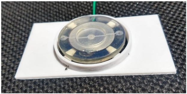
One of our 3D printed microfluidic devices.
2.1 Acoustofluidic microplate
We are developing microtiter plates with microfluidics technology. One advantage is the low consumption of reagents in a controlled microenvironment. This is one of the selected projects of Catalisa/Sebrae.
Giclenio C. Silva & Glauber T. Silva
2.2 Acoustofluidic micromixer
Giclenio C. Silva & Glauber T. Silva
2.3 Measurement of cell density and compressibility
Harrisson Santos & Glauber T. Silva
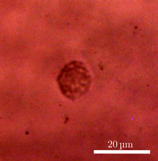
A levitating macrophage ready to be weighted in an acoustofluidic device.
2.4 Acoustofluidics for leishmaniasis diagnostic (PPSUS 2021/FAPEAL)
Aline C. Queiroz & Glauber T. Silva
3. Raman-acoustofluidics
Inside a microwell, cells and other analytes are acoustically levitated and concentrated, allowing for an enhanced Raman spectroscopy.
3.1 Monitoring infection assays of Leishmania
Harrisson Santos, Ueslen Rocha, Magna S. Alexandre-Moreira & Glauber T. Silva
Publications:
HDA Santos et al., Raman-acoustofluidic integrated system for cell analysis, submitted, 2021.
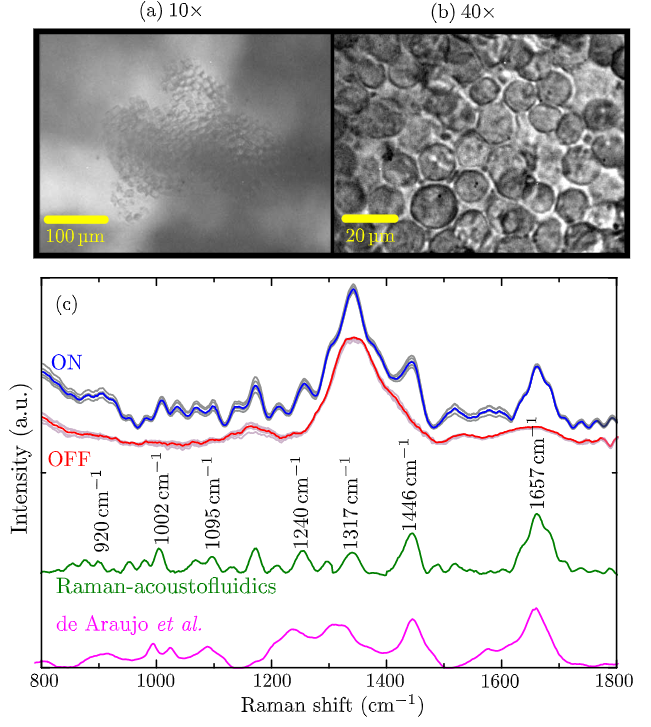
Bright-field images of macrophages in the levitation plane taken by (a) a 10× and (b) a 40× objective lens. (c) The room-temperature Raman spectra (gray lines) of a single macrophage. The red and blue curves correspond to the average spectrum with the device ‘OFF’ and ‘ON’, respectively. The green curve is the averaged Raman spectrum after background subtraction. For comparison, the spectrum obtained from fixed macrophages is also shown (pink curve).
3.2 Biochemical analysis of red blood cells
Ueslen Rocha, Ana C. Leite & Glauber T. Silva
Collaboration with IQB-UFAL.
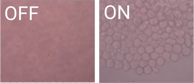
Red blood cells trapped in the acoustofluidic device. This configuration is suitable for Raman spectroscopy.
5. Ultrasound subwavelength imaging
The purpose of this project is to image structures and tissue with ultrasonic beams that beat the diffraction limit.
J. P. Leão-Neto, J. H, Lopes & Glauber T. Silva
Publications:
- JP Leão-Neto et al., Subwavelength focusing beam and superresolution ultrasonic imaging using a core-shell lens, Physical Review Applied 13, 014062, 2020.
- EB Lima, VHS Santos, AL Baggio, JH Lopes, JP Leão-Neto, GT Silva, An image formation model for ultrasound superresolution using a polymer ball lens, Applied Acoustics 170, 107494, 2020.
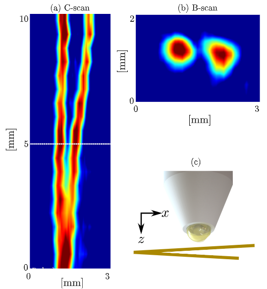
Ultrasound superresolution image of a copper wire forming a Y-intersection (with a diameter of a human hair) and immersed in water at room temperature. (a) The transverse (C-scan) image planes at the transducer focal plane. (b) B-scan image corresponding to a plane at 4.7 mm away from the transducer. (c) Transducer scanning the wire.
6. Automation for microfluidics (micro-automation)
We develop and fabricate control units for microfluidic devices such as pumps, temperature cells, etc.
Ícaro Bezerra & Glauber T. Silva
7. Cell mechanobiology with acoustofluidics
Ultrasonic waves and shear flows can deform soft mater including biological cells. We are developing acoustofluidic chips to deform cells and provide a readout of the cell membrane elasticity.
7.1 Acoustic deformation of giant unilamellar vesicles
Glauber T. Silva
Collaboration with the University of Bristol (Newton Fund)
Publications:
GT Silva et al., Acoustic deformation for the extraction of mechanical properties of lipid vesicle populations, Physical Review E 99, 063002, 2019.
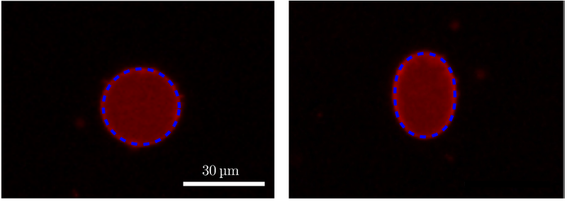
(Left) A trapped vesicle without deformation. (Right) Acoustically deformed vesicle with aspect ratio 1.33. The blue-dashed contour is our theoretical prediction.
Sponsors


